Scientific Discussion of Plantar Fasciitis
What is the etiology of Plantar Fasciitis?
Scientific Discussion of Plantar Fasciitis
What are the risk factors that lead to Plantar Fasciitis?
Scientific Discussion of Plantar Fasciitis
Scientific Discussion of Plantar Fasciitis What’s the disease progression of Plantar Fasciitis?
Previous
Next

A Scientific Discussion of Plantar Fasciitis

Plantar fasciitis (also referred to as subcalcaneal pain, painful heel syndrome, calcaneodynia, heel spur syndrome, runner’s heel, and calcaneal periostitis) is the most frequently reported cause of heel pain.  It involves pain and inflammation of a thick band of tissue, called the plantar fascia that runs across the bottom of your foot and connects your heel bone to your toes. In untreated or poorly managed cases, Plantar Fasciitis can dramatically impact physical mobility and overall quality of life.

Plantar Fasciitis is fairly common:

  • 9% of all running injuries in the United States are due to plantar fasciitis (1).
  • 15% of all the reported cases of foot pain are due to plantar fasciitis(2).
  • The estimated lifetime risk of developing plantar fasciitis is about 10% in US population(3).
  • More than 2 million individuals are treated in the United States each year for Plantar Fasscitis related pain and discomfort(4).
  • The economic burden of plantar fasciitis is estimated to be between $192 to $376 million per annum in United States(5).

Unfortunately, the exact cause or pathophysiology of Plantar Fasciitis is not known in about 85% cases; but it is believed that the condition is mostly multifactorial in origin i.e. a wide variety of mechanical, anatomical and environmental factors plays a vital role in its pathogenesis(5). Although Plantar Fasciitis is more common in dynamic individuals; the risk is significantly high in general population as well, especially middle-aged women with a sedentary lifestyle(6).

Anatomy of the Plantar Fascia

The structural integrity of foot is reinforced by thick bands of connective tissues that form plantar arches and fascia in order to enhance biomechanical alignment and optimal physiological functioning of the foot to promote effortless walking and weight bearing.

The Plantar Fascia consists of thick connective tissue which supports the Medial Longitudinal Arch on the sole (plantar side) of the foot. It runs from the medial tuberosity of the calcaneus (the inside of the heel bone) forward to the heads of the metatarsal bones. The plantar fascia is made up of predominantly longitudinally oriented collagen fibers and it looks like a white, flattened or ribbon-like tendinous expansion. it includes a thick central component and thinner medial and lateral components. Contributes to the support of the foot by absorbing as much as 14% of the total load of the foot.

 Risk Factors

There are significant risk factors that plays a vital role in the initiation and progression of plantar fasciitis:

  • Weight gain or other metabolic disorders Obesity aggravates the cartilage loss and connective tissue degeneration that weakens the support and strength of connective tissues of the foot. All these factors eventually affect the integrity of the plantar arches and leads to aggressive wear and tear related damage. In addition, diabetics are also at a fairly higher risk of experiencing plantar fasciitis due to atrophy of intrinsic muscles and peripheral motor neuropathy. A higher body mass index (>30 g/m2) can increase the risk of developing Plantar Fasciitis by 5.6%(7).
  • Certain occupation that requires vigorous lower limb motion/ activity: Some examples include; professional athletes (soccer, basketball, volleyball) as well as occupations that requires long hours of standing or walking (such as soldiers and teachers) are at high risk of developing this musculoskeletal pathology. Research indicates that overuse injury is the most common cause of plantar fasciitis in athletes owing to training errors (use of worn out shoes, practice of unyielding surface and improper foot gear).
  • Anatomical variations: Congenital or acquired deformities of foot like claw toes, flat foot syndrome, fallen arches, prominent metatarsal heads, and excessive pronation of foot (associated with 86% cases of Plantar Fasciitis), can also aggravate the risk of developing plantar fasciitis and other foot pathologies due to altered biomechanics and gait changes. Likewise, more than 50% patients with Plantar Fasciitis have heel spurs(5).
  • Overexertion: Poor conditioning, improper training and inadequate warm-up before engaging in vigorous foot activity (such as running, sports training etc.) is one of the leading risk factors that may aggravate the risk of other foot disorders as well. Study suggested that individuals with highly dynamic lifestyles (people who spend major part of their day on foot) are 3.6 times more likely to develop plantar fasciitis than control group(8).
  • Inadequate footwear: Ill-fitting, worn-out shoes affects the natural biomechanics of the foot and affects the balance and coordination.
  • Poor biomechanics: Certain biomechanical factors such as poor range of ankle dorsiflexion (< 10 degrees) is associated with 2.1% higher risk of developing Plantar Fasciitis(8). According to another study, angle of dorsiflexion ≤0° is associated with 23.3% higher risk of developing plantar fasciitis(7). Other biomechanical risk factors include excessive femoral anteversion, excessive lateral tibial torsion and leg length discrepancy,
  • Abnormal weight distribution due to above listed factors (in isolation or combination) can further aggravates the degeneration process.

Etiology:

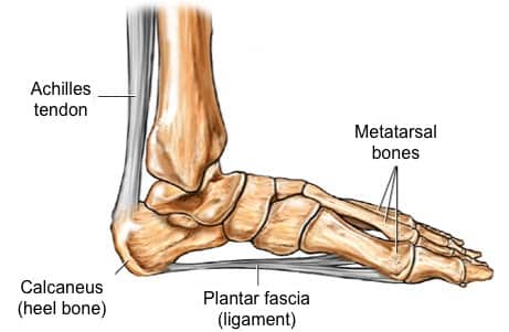
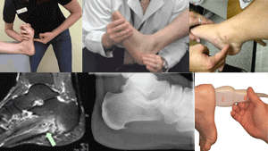
Plantar fasciitis is the most frequently reported and disabling disorder of the foot, but very little is known about the exact cause. It is thought that ongoing injury which causes microtearing and persistent inflammation of the plantar fascia at its origin (i.e. calcaneus) resulting in the degeneration of the connective tissue band(9).

It has been suggested that plantar fasciitis represents a form of tennis elbow at the heel with the condition being caused by repetitive microtrauma at the point of insertion of the plantar fascia.

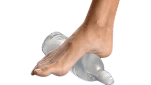
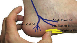
Whenever the heel is in contact with the ground (such as during walking), the foot automatically pronates due to inward movement of tibia. This leads to stretching of the plantar fascia and the pressure generated due to this stretching flattens the plantar arches. In simple words, this arrangement acts like shock absorbers in accommodating the pressure and stress from walking. In the presence of certain contributing risk factors (listed above such as obesity, improper foot gear etc.), the fibers of plantar fascia undergo excessive stretching and micro tearing(1)(10). Most commonly, the micro-tearing begins at the site of origin of Plantar Fasciitis – the medial prominence or tuberosity of the calcaneus. If left untreated, the process of tearing causes degeneration of fascial fibers and associated connective tissue elements.

Pathogenesis / mechanism of injury

The plantar fascia act as a shock-absorber for the foot (like a bowstring) supporting the foot arches and musculoskeletal framework of the foot. However, with persistent exposure to undue stress, tension and pressure, your plantar fascia undergoes excessive wear and tear related damage. Plantar fasciitis probably begins with small tears that occur during activity and that the body usually repairs. But with repeated stress and subsequent ongoing degeneration, these tears accumulate deceasing the strength and stability of plantar fascia and also makes it more vulnerable to further strain and stress injuries(11). Most individuals develop unconscious changes in gait and posture to minimize the pain; however, this further aggravates the tissue damage and widespread biomechanical disruption and subsequently leads to other problems such as ankle pain, knee osteoarthritis, back discomfort, pelvic instability, etc.

Disease Progression

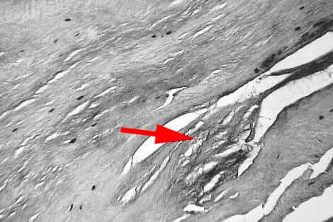
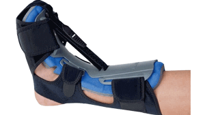
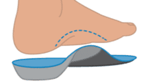
Plantar fasciitis is usually a self-resolving condition especially when treated early with conservative treatments. However, once the degeneration and persistent inflammatory reaction sets in, it is usually a downhill course. The plantar fasciitis usually presents with persistent pain in the infero-medial aspect of the heel. The pathophysiology of pain is complex and is often described as a multitude of various pathological processes such as loss of normal elasticity of fascial fibers, abnormal changes in the vascularity due to fibrosis, thickening of the plantar fascia and inflammatory destruction. In the absence of any meaningful interventions to control the process or minimize the ongoing damage (stretching, orthotics, shoes modification, night splinting etc.), most patients develop severe morbidity and disability.

Histology

Healthy fescia
Plantar Fascia degeneration in Plantar Fasciitis

The histological changes in plantar fasciitis vary according to the age of the patient and duration of symptoms. The Plantar Fascia fibers are thicker in younger people and as an individual ages, the concentration and continuation of fibers decrease(12).

Most investigators prefer the term fasciosis to define the degenerative pathological processes in the Plantar Fasciitis instead of fasciitis to indicate that plantar fibrosis is a degenerative process without inflammation. Histological examination of plantar fasciitis reveals myxoid degeneration of the fascia(13) associated with fragmentation. Severe and chronic cases of plantar fasciitis show typical granulomatous changes besides localized fibrosis and scarring(14). Other histological findings may include collagen necrosis, angiofibroblastic hyperplasia, chondroid metaplasia and matrix calcification, scar deposition etc.)

In view of such findings, it is  surprising that so many patients seem to respond to local steroid injections and oral anti-inflammatory drugs. However, this paradox is hardly unique to plantar fasciitis. Both lateral and medial epicondylitis respond to similar treatments in spite of the inability of modern histologists to find inflammatory changes at the site of tenderness. It must also be remembered that histology is not obtained on all patients with heel pain and the absence of inflammation in the subgroup (less than 1%) that comes to surgery may not be an accurate representation of plantar fasciitis patients as a whole.

References
  1. Fasciitis, P. (2005). Plantar fasciitis: diagnosis and therapeutic considerations. Alternative medicine review, 10(2), 83-93.
  2. Crawford, F., & Thomson, C. E. (2003). Interventions for treating plantar heel pain. The Cochrane Library.
  3. Cornwall MW. McPoil TG. Plantar fasciitis: etiology and trciitnient. J Orthop Sports Pins Tlicr l999;29:756-76.
  4. Tong, K. B., & Furia, J. (2010). Economic burden of plantar fasciitis treatment in the United States. Physical therapy, 8(4), 11.
  5. Crawford, F., & Thomson, C. E. (2003). Interventions for treating plantar heel pain. The Cochrane Library.
  6. Pfeffer, G., Bacchetti, P., Deland, J., Lewis, A. I., Anderson, R., Davis, W., et al. (1999). Comparison of custom and prefabricated orthoses in the initial treatment of proximal plantar fasciitis. Foot & Ankle International, 20(4), 214-221.
  7. Fasciitis, P. (2005). Plantar fasciitis: diagnosis and therapeutic considerations. Alternative medicine review, 10(2), 83-93.
  8. Riddle, D. L., Pulisic, M., Pidcoe, P., & Johnson, R. E. (2003). Risk factors for plantar fasciitis: a matched case-control study. The Journal of Bone & Joint Surgery, 85(5), 872-877.
  9. Soysa, A., Hiller, C., Refshauge, K., & Burns, J. (2012). Importance and challenges of measuring intrinsic foot muscle strength. J Foot Ankle Res, 5(1), 29.
  10. Wearing, S. C., Smeathers, J. E., Urry, S. R., Hennig, E. M., & Hills, A. P. (2006). The pathomechanics of plantar fasciitis. Sports Medicine, 36(7), 585-611.
  11. Rome, K., Howe, T., & Haslock, I. (2001). Risk factors associated with the development of plantar heel pain in athletes. The Foot, 11(3), 119-125.
  12. Cole, C., Seto, C., & Gazewood, J. (2005). Plantar fasciitis: evidence-based review of diagnosis and therapy. Am Fam Physician, 72(11), 2237-42.
  13. Snow, S. W., Bohne, W. H., DiCarlo, E., & Chang, V. K. (1995). Anatomy of the Achilles tendon and plantar fascia in relation to the calcaneus in various age groups. Foot & ankle international, 16(7), 418-421
  14. Schepsis, A. A., Leach, R. E., & Gouyca, J. (1991). Plantar fasciitis: Etiology, treatment, surgical results, and review of the literature. Clinical orthopaedics and related research, 266, 185-196.
  15. DeMaio M, Paine R. Mangine RE. Dre/D Jr. Plantar fasciitis. Orthopedics 1993:16:1153-1163.
Request Appointment >


Let’s Talk!

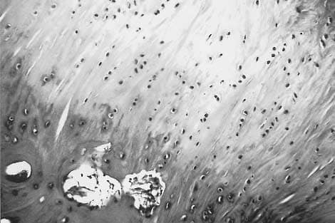
We’re here to help you with any questions you may have. Please don’t hesitate to reach out to us.

  • This field is for validation purposes and should be left unchanged.